Get How To Draw Heart Diagram Class 10 Cbse Images. Not everyone is an artist so some class 10 board exam aspirants must be dealing with a lot of trouble for their diagrams. Solution of life processes page no 74 part 2 biology classes events the chattery.
Human Heart Diagram Class 10th ~ DIAGRAM from i.pinimg.com Human heart diagram science diagram biology diagram class 10 science diagram #humanheartdiagram #humanheartdrawing. How to draw human heart step by step in very easy way for class 10 students taught by fine arts guruji to draw human heart. Well, you are in the right place as this article will help you find the.
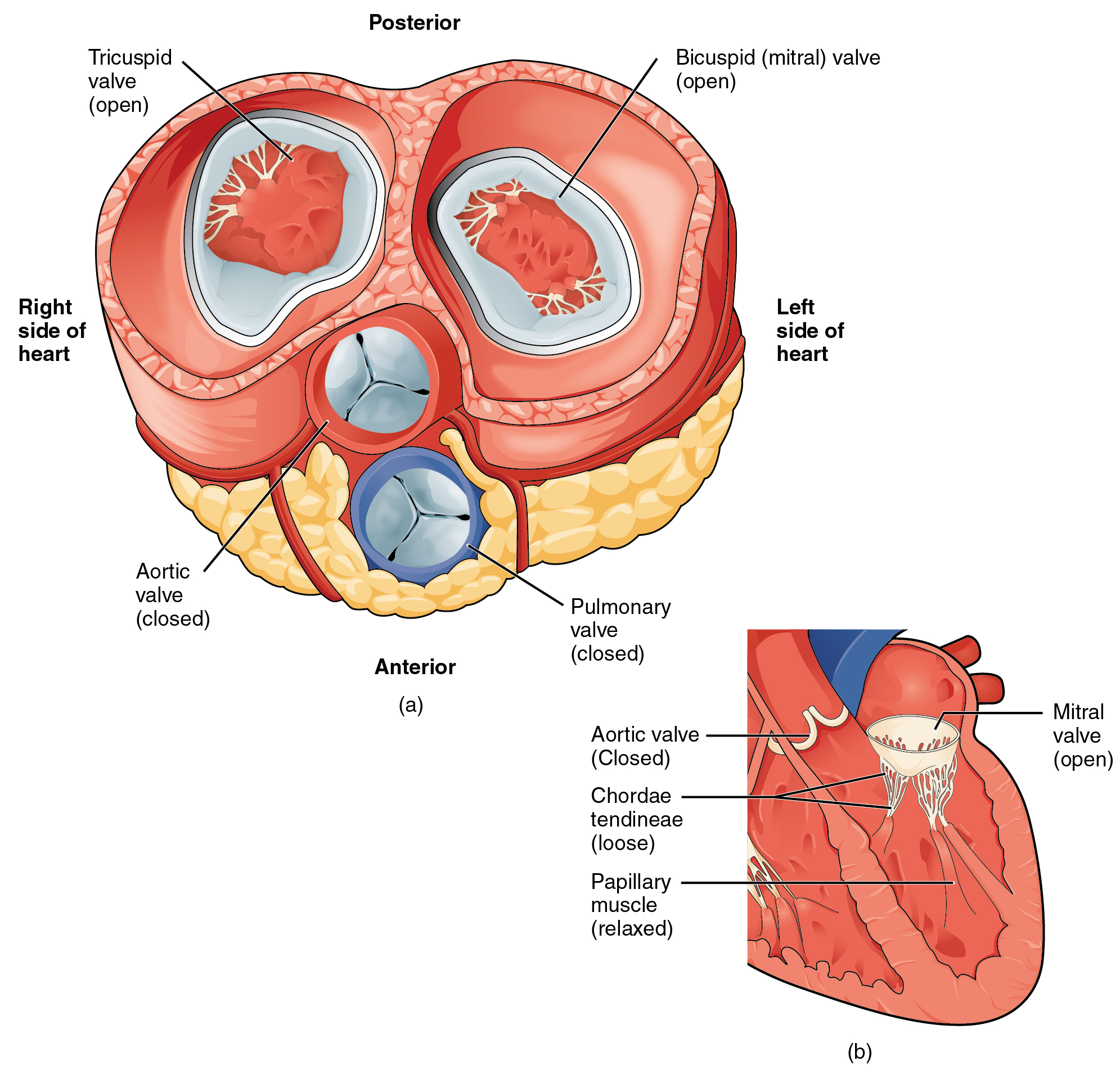
How to draw a heart diagram?
At least 30 minutes a day, or three and a half hours per week. Draw the lower half of an acorn shape so it's tilted to the left. Wish you best of luck. How can i draw a diagram of the human heart and label its different parts?
41+ Heart Diagram Labeled Drawing Background. 1024x1041 heart anatomy drawing with label the human heart diagram labeled. Daniel nelson on january 1, 2019 1 comment.
File:Diagram of the human heart (cropped) de V2.svg ... from upload.wikimedia.org Labeled drawing of the heart at getdrawings free for personal. Make an oval that touches each point of the. The function of heart is quite complex.
1200 x 800 jpeg 159 кб.
The easiest way to draw a heart diagram is using edrawmax by simply choosing a template, then export it there are two ways to draw a heart diagram, one is from the sketch which could spend a lot of time for creating. This digital heart diagram is a simple and clearly drawn template. Draw internal structure of heart. Label heart anatomy diagram printout printable cashewapp co.
32+ What Are The Names Of The Parts Of The Heart Background. The upper two chambers are called atria (singular: For this reason, cardiac muscle has an extensive network of blood vessels to bring oxygen to the contracting cells and to remove waste products.
6 1 Human Heart Parts - YouTube from i.ytimg.com For this reason, cardiac muscle has an extensive network of blood vessels to bring oxygen to the contracting cells and to remove waste products. The heart beats continuously, pumping the equivalent of more than 14,000 litres of blood every day through five main types of blood vessels: The four valves of the heart all keep blood moving correctly.
I am going to list technical terms and names of various parts of the heart on the screen, but i so what is going to happen is these upper chambers, which i have listed as 1 and 3 are going to fill with blood, chamber 1 is going to fill with deoxygenated.
What is the name of the substance that 14. Changes of pressure allow the blood to flow through the cardiac cycle. If one of your organs is working that hard, it makes sense to learn about how it functions! The red blood cells are entering the chambers of the heart the part that they're going to see is going to be the endocardium so this is what it looks like and this is that green layer all the way around that i've drawn now so if i was to draw it.
View Heart Diagram Pulmonary Artery Background. The pulmonary artery, or pulmonary trunk, originates from the bottom of the right ventricle of the heart. The pulmonary artery, being an exception, carries deoxygenated blood to the lungs for purification.
Chapter 42: Circulation and Gas Exchange. - AP Biology ... from classconnection.s3.amazonaws.com In this article we will consider the structure and anatomical relationships of the aorta, pulmonary arteries and veins, and the superior and inferior vena cavae. The veins carry impure blood from different parts of the body to the heart for oxygenation. Contraction of the heart pumps blood through the heart and out into arteries.
The oxygen present in the tiny air sacs.
In this image, you will find superior vena cava, right pulmonary artery, right pulmonary veins, right atrium, inferior vena cava, coronary sinus, right coronary artery, coronary sulcus, posterior interventricular artery in it. For blood to pass through the heart from the left atrium to the left ventricle, it must first pass through a) an artery c) a vein b) a valve d) a capillary Small branches dive into the heart muscle to bring it blood. The pulmonary artery, being an exception, carries deoxygenated blood to the lungs for purification.
28+ Heart Vessels Anatomy Diagram Background. The great vessels that originate from the heart, radiate their branches to the head and neck, the thorax and abdomen and the upper and lower limbs. In this article we will consider the structure and anatomical relationships of the aorta, pulmonary arteries and veins, and the superior and inferior vena cavae.
Heart Anatomy · Anatomy and Physiology from philschatz.com Simply choose the topic you need to revise. Don't forget to share this picture with others via facebook, twitter, pinterest or other social medias! The aorta sends oxygenated blood from the heart to the entire body.
Explore the fine layers and features of the vessels like never before.
Fetal circulation diagram blood flow. Major blood vessels of the heart diagram unique heart anatomy. It is located in the middle cavity of the chest, between the lungs. Michael servetus research study on the manuscript of paris by servetus (1546 description of the pulmonary circulation).

_de_V2.svg/1024px-Diagram_of_the_human_heart_(cropped)_de_V2.svg.png)